

Latest Trend in Biomedical Instrumentation:
DIGITAL MAMMOGRAPHY
Do you wish to join the research?
Potential Researchers Welcome.
DIGITAL MAMMOGRAPHY
Do you wish to join the research?
Potential Researchers Welcome.
Ultra Sonic Based Phosphor Filled Balloons For Breast Density Normalization (ULBDN)
The normal X raying of breasts for mammography suffers from non-uniform x ray image contrast due to the oval shape of the breast on compression and the corresponding uneven X-ray attenuation due to which the fed x-ray power tends to create an effect of over exposed, under exposed and normally exposed regions in the x-ray image (see Fig). This uneven contrast constraints the use of the obtained x-ray image for identifying the micro-calcifications and there by leading to false identifications. Thus, it is a requirement that the breast surface is normalized such that the x-ray attenuation at any part of the breast is the same there by obtaining no contrast gradient owing to the compressed breast shape.
THE PROPOSED METHOD:
(NOT YET DISCLOSED)
(NOT YET DISCLOSED)
RESULTS:
We conducted a series of x-ray image acquisition using different chemicals like lead compounds, bismuth compounds, phosphor compounds etc. Out of all these chemicals, xxx is best suiting for the application. The results showing the difference between ordinary mammogram and a mammogram that acquired using ULBDN technique are illustrated below.
Fig.1
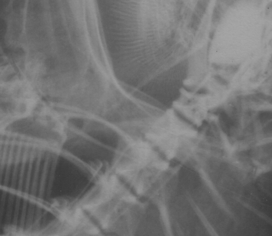
X-ray image of head portion of a fish acquired in conventional method.
X-ray image of head portion of a fish acquired in conventional method.
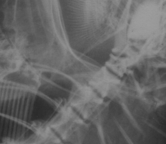
Figures 1 & 2 show the head section of the fish which is a denser portion and thus the image remains the same even after the normalization process.
Fig.2
X-ray image of head portion of a fish acquired in ULBDN method.
X-ray image of head portion of a fish acquired in ULBDN method.
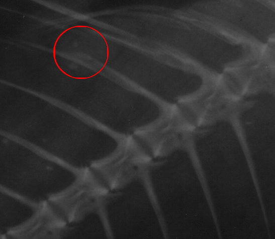
The Figures 3 & 4 show the tail section, which is a lower density and thus the image in Fig. 4 is a contrast enhanced version of that of Fig .3 after the ULBDN technique is applied. It can be clearly seen from the circled area in Fig.4 that shows the X-ray attenuating calcium compounds that are unseen in Fig.3.
Fig.3
X-ray image of tail portion of a fish acquired in conventional method.
X-ray image of tail portion of a fish acquired in conventional method.
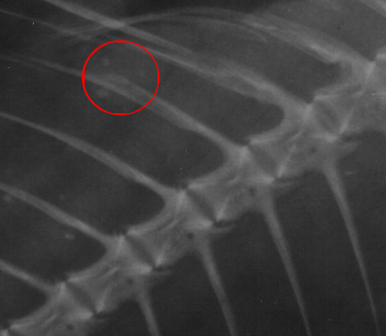
Fig.4
X-ray image of tail portion of a fish acquired in ULBDN method.
X-ray image of tail portion of a fish acquired in ULBDN method.
REFERENCES:
1."The Breast", Stephen A Feig and Catherine W Piccoli.
2. Feig S A 1987 Mammography equipment: principles, features, selection. Radiol CCClin North Am 25:879-911.
3. Sickles E A 1988 Practical Solutions to Common Mammographic Problems : tailoring the examination. ARJ 151:31-39.
4. Kopans D B , Meyer J E , Homer M J ,Grabble J 1983 Dermal deposits mistaken for Breast calcification .Radiology 149 :592-594.
5. Tabar L , Dean P B 1983 Teaching Atlas of Mammography . Thieme , Newyork. Sabel M, Aichinger H Recent developments in breast imaging . phy med boil.1996; 41:315-
www.electrogenes.com