

Latest Trend in Biomedical Instrumentation:
DIGITAL MAMMOGRAPHY
Do you wish to join the research?
Potential Researchers Welcome.
DIGITAL MAMMOGRAPHY
Do you wish to join the research?
Potential Researchers Welcome.
OUR TEAM
DESIGNER - TONY GLADVIN GEORGE
CO DESIGNER - RAJESH.S
SUPPORT - Prof. Dr. A.NAGAPPAN
CO DESIGNER - RAJESH.S
SUPPORT - Prof. Dr. A.NAGAPPAN
INTRODUCTION
Digital mammography is a latest technique of acquiring the x-ray image of the mammary gland in digital format. It is used for the diagnosing of cancer. According to the recent surveys held in India, the mortality rates due to the mammary gland cancer is more than 5000 per year. The only cure is to remove the cancerous tissues in the early stages of its growth. Unluckily, the mammography units available in India are not up to the mark for a 100% successful diagnosis. Here comes the role of Digital Mammography.
Digital mammography has several advantages over conventional methods. Standard screen-film systems currently used in mammography have a number of well-known limitations including poor detective quantum efficiency and limited dynamic range. Digital mammography incorporates high performance digital detectors to replace these systems and allow more direct utilization of Digital Processing Techniques, 3D Reconstruction Techniques and Computer-Aided Diagnosis in the detection of subtle mammary gland cancers. The detector is comprised of an array of modules, each of which is an independent charge coupled device (CCD) sensor that is fiber optically coupled to an x-ray-to-light converter (ASNCS).
Early detection is crucial in treating mammary gland cancer. By offering increased contrast and resolution, a wider dynamic range, a higher signal-to-noise ratio, and real-time image enhancement capabilities, digital mammography will provide important improvements in early detection rates and could ultimately help save lives.
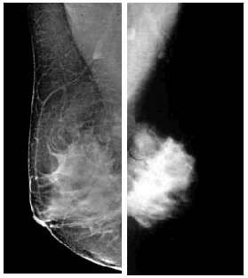
Image acquired using Digital detector in Digital Mammogram
Image acquired using Screen Film Mammogram
A digital X-ray detector technology first developed for use by structural biologist is now being used to produce speedy, high-resolution digital images of breast tissue that could replace standard mammograms as a screening technique. The images produced from our unit, shows much better detail and resolution compared to the standard mammogram. Better quality images could lead to earlier detection of changes in tissue that might indicate cancer. About 20 percent of breast tumors are missed by conventional mammograms, and the difficulty of detecting them increases in younger women whose tissue is denser. When tumors are found at the earliest stage of growth, the survival rate is close to 100 percent.
The digital X-ray detector designed is based on CCD arrays. The (ULBDN*) technique developed is used for the normalization of breast density which eliminates the contrast gradient owing to the compressed breast shape. The "Anti Scattering Nano Chemical Screen (ASNCS*)" technique is used for removal of distortions of the image due to scattering of X-rays, resulting in better quality images. The detector captures the 2-dimensional X-ray image of the breast at two inclinations separated mutually by an angle of 90° in the same breast plane*. Finally, the images obtained on the CCD - arrays are interfaced to the computer and by using the mathematical transformations, the 3-dimensional breast image is reconstructed from the two 2-Dimensional images thus obtained , which aids in accurate computer aided diagnosis and robotic biopsies*.
(* - our contribution)
(* - our contribution)
In a digital mammography system the film/screen cassette is replaced by a phosphor screen, which detects the x-ray photons leaving the breast. The phosphor screen converts the x-ray photons into light, which is transferred through a fiber optical reducer to the CCD (Charged Coupled Device) detector. The matrix of a typical CCD is 1600 by 1600; therefore each pixel can resolve 0.05mm. The CCD converts the incident light into a digitized analogue signal, which is a pair of 2D image. These 2D images are reconstructed into 3D and this is displayed onto a computer monitor. Rather than reaching an optical density of 1.4, the exposure is terminated when a sufficient signal to noise ratio is achieved. A digital mammogram's "dynamic range" - essentially a measure of the range of shades from black to white - is about 10,000, compared to only about 100 for a film mammogram.
FEATURES OF THE DIGITAL MAMMOGRAPHIC UNIT UNDER DEVELOPMENT
Image size: 18.6 x 24.8 cm
Detector design: Array of 12 CCDs (3 x 4)
Detector dimensions: 30 cm (w), 36 cm (d), 11 cm (h)
Phosphor material: Caesium Iodide
CCD to phosphor coupling: Fiber optic bundle with demagnification factor of ~2.2 (manufacturer's literature)
Pixel array (per CCD): 1600 x 1600
Pixel array (total): 4800 x 6400
References:
1) Kalata, K., Phillips, W.C, Stanton, M., and Li, Y. (1990). "Development of a Synchrotron CCD-Based Area Detector for Structural Biology ", Proc. Soc. Photo-Opt. Instr. Eng. 1345 270-280.
2 )Strauss, M.G., Westbrook, E.M., Naday, I, Coleman, T.A., Westbrook, M.L., Travis, D.J., Sweet, R.M., Pflugrath, J.W., and Stanton, M. (1990). "Large aperture CCD X-ray detector for protein crystallography using a fiberoptic taper", Proc. Soc. Photo-Opt. Instr. Eng. 1447 12-27.
3) Stanton, M, Phillips, W., Li, Y. and Kalata, K. (1992) "The Detective Quantum Efficiency of CCD and Vidicon Detectors for X-ray Crystallographic Applications", J. Appl. Cryst. 25, 638-645.
4) O'Mara, D., Phillips, W., Stanton, M., Sarof, D., Naday, I. and Westbrook, E. (1992) "Design Criteria and Development of Components for a Modular CCD-based Detector for X-ray Crystallography",, Proc. Soc. Photo-Opt. Instr. Eng.1656, 450-456.
5) Kalata, K., Stanton, M. and Phillips, W.C. (1992). "The Detective Quantum Efficiency of Television Area X-Ray Detectors", J. X-Ray Sciences and
Websites:
1) www.rose.brandeis.edu
2) www.epanorama.net
5) www.biomechs.net
www.electrogenes.com